There are two main parts to the Respiratory System:
1. The Upper Respiratory Tract 2. The Lower Respiratory Tract The Upper Respiratory Tract consists of the mouth, nose (including sinuses and smell), nasopharynx, oropharynx, laryngopharynx, and larynx. These are mainly all located in the throat area and above. The Lower Respiratory Tract includes the trachea and the lungs. The purpose of the Respiratory System is to take oxygen into our bodies and release carbon dioxide. It does this by inhaling the oxygen through our nose or mouth (the best way to inhale is through the nose because of the cilia or little hairs that are all throughout it that help filter the air taken in and moisten it with mucus for the best possible intake but the mouth also works as it is shorter and wider so more air can be taken in at once). Then the air is taken to our lungs where it is distributed by the bronchi to the red blood cells which carry it through the body until the carbon dioxide is brought back to the lungs where it is then exhaled out of the body. Terms of the Respiratory SystemMouth (oral cavity): The external opening inferior to the nose as it does not filter and moisten the air taken in as well as the nose does. The mouth is used most importantly when trying to catch one's breath and the mouth's shorter pathway to the lungs works better to get the air in faster. This was shown as the bike pump's air hose in our model.
Nose (nasal cavity): Main external opening, has cilia that help filter and moisten the air inhaled. Larynx: Also known as the voice box. It's also used as a defense mechanism in case any food was to pass into the esophagus when swallowing. In the larynx is the Arytenoid cartilage, Corniculate cartilage, Cricoid cartilage, Cricothyroid joint, Epiglottis, Thyroid membrane, and Thyroid cartilage. Trachea: Brings air to and from the lungs. This was shown as the main tube of the bike pump. Lungs: The part of the Respiratory System that allows oxygen to enter the bloodstream and that also lets carbon dioxide leave the body. This is shown as the base of the bike pump. Alveoli: The tiny air sac in the lungs. Bronchi: Conducts air into the lungs. Below is our slideshow that explains all of the processes and how they work together! Also please take a look at some of my work in English! Link below! |
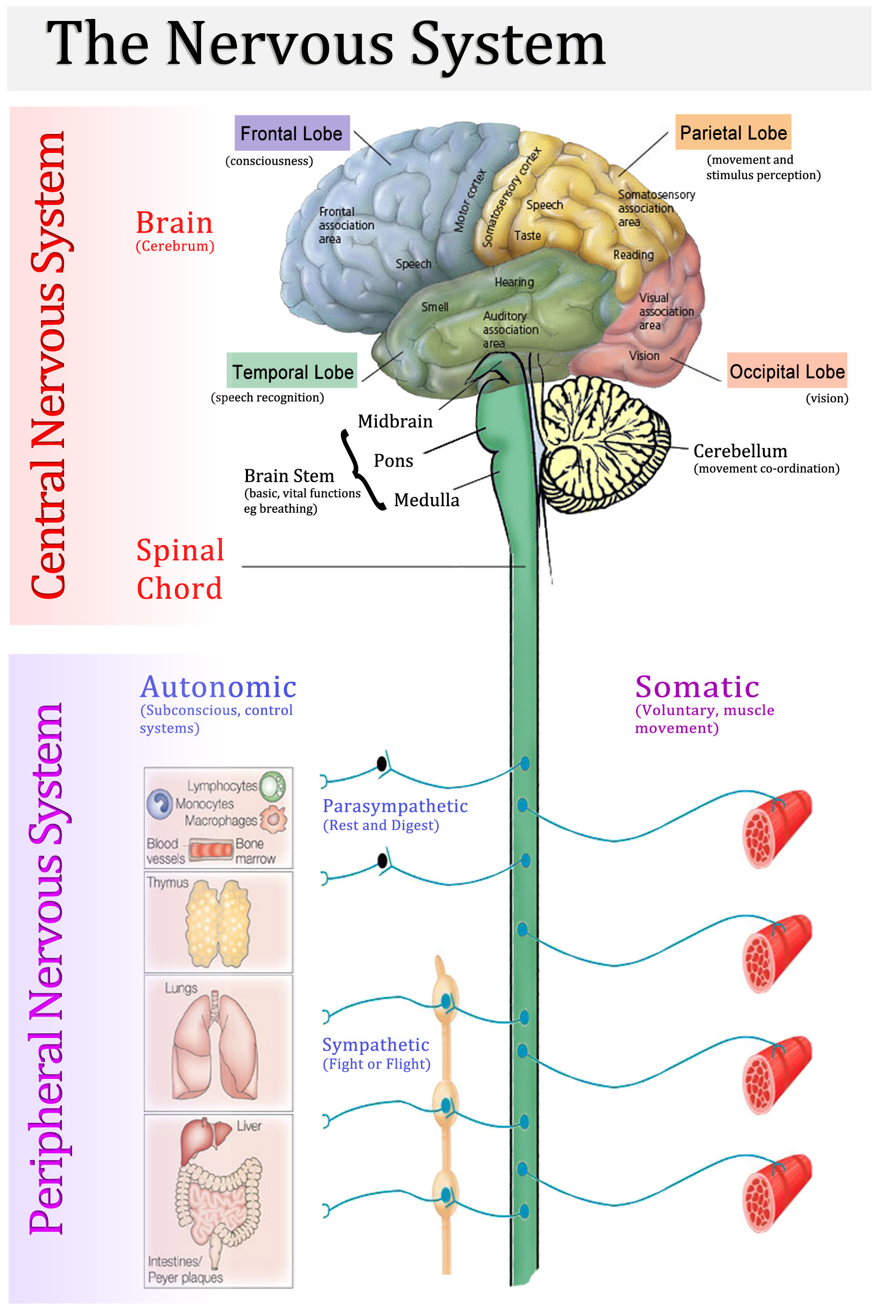
The nervous system consists of the brain, spinal cord, sensory organs, and all nerves that connect to the rest of the body. The main purpose of the nervous system is to send messages from the brain to the rest of the body. It is split into two main parts - the Central Nervous System and the Peripheral Nervous System. The Central Nervous System icludes the brain, which has four main parts, the first part being the Cerebrum which is made up of its own 4 parts- the Frontal Lobe, Parietal Lobe, Temporal Lobe, and Occipital Lobe. The second part of the brain is the Cerebellum which is located behind and below the Cerebrum. The third part of the brain is the Dicephalon which consists of the Thalamus, and the Hypothalamus. The fourth part is the Brain Stem which is located between the pons and the spinal cord, and is only about 1 inch long.
Terms of the Nervous SystemNerves: Cyllindrical bundles of fibers which start at the brain and central cord and extend to just about every part of the body. The brain uses nerves to send signal impulses all around the body so that you can function. This was represented byt the two slinkies in our model.
The Autonomous Nervous System: Regulates specific body processes like the rate of breathing and blood pressure - things that seemingly take no effort to do because they happen automatically because of your nervous system. This was represented by our model as a whole, as we portrayed the process of breathing, which is automatic. Frontal Lobe: Performs functions like expressive language, reasoning, and the ability for you to drive a car.Damage to this lobe ca cause a lower level of sociability, sexual habits, attention, and more. Parietal Lobe: Processes the information that the brain recieves from physical aspects like pain, touch, and pressure. Any damage to this lobe and it could cause language problems, and loss of verbal memory. Occipital Lobe: Interprets the information recieved from the eyes and is located near the back of the brain. Any damage to this lobe and your vision could be impaired. Temporal Lobe: Forms memories and processes sound taken in by the ears and is located at the bottom of the brain. Any damage to this lobe and it creates problems with language, speech perception, and memory. Neurons: A specialized cell that transmits nerve impulses. In our model this was represented as the beads in the Newton's Cradle. Spinal Cord: The cyllindrical bundle of nerve fibers and tissue enclosed in the spine and reaches out to just about every part of the body. Brain: The main organ of the Nervous System that is made up of soft nervous tissue enclosed into the skull. This was the water bottle in our model. The Respiratory System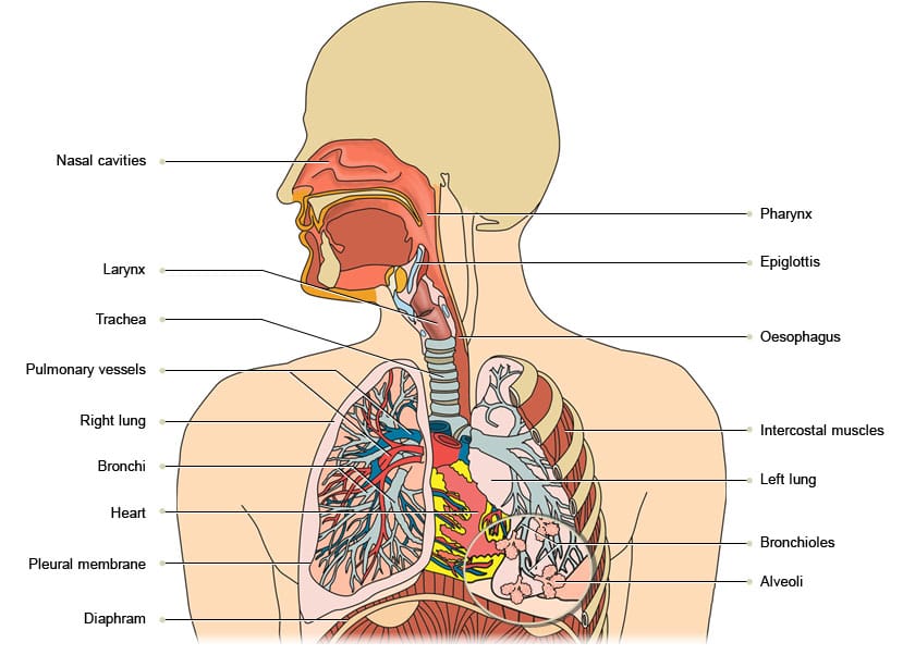
The Relationship Between the Nervous System and the Respiratory System
|
Reflection:
Overall, this project expanded my knowledge in a variety of ways. This was the first project since freshman year that we got to build something, and that was really exciting. It really made me learn in a whole new way because you aren't just writing things down, but building them as well. A challenge we faced during this project was finding something that related to the two systems. It was difficult because our two things didn't quite go together (for the first full day of the project I thought we had the circulatory and respiratory systems so I researched the circulatory a lot until I finally realized it was the wrong one). Once we finally found a relation- the nervous system sending signals to the lungs to breathe and regulating the size of the air pipes, it was very hard to find a way to model that. Eventually we found our solution to be slinkies and a bike pump! These all fit perfectly with what we needed to do. However, this brought up another problem- how do we show the difference between the inhaling and exhaling and the two different nerve signals that are sent to do these processes? It took a lot of imagination, but we ended up painting one half of the bike pump's tube blue to symbolize the oxygen being inhaled, and the other side, which was left black, would symbolize the carbon being exhaled. During the demonstration we use the blue side facing front and pulled the handle up to inhale, and spun it halfway to the blackside and pushed down to show the exhale. Then, we had two separate slinkies attached to either side of the brain which each connected to one side of the bike pump's handle. Fiinally, we had found a great representation model! This was a great project and great group to work with!
